I am grateful to a friend for asking me to look at the ketamine and medetomidine used during the recent surgery of her 8 month old Burmese kitten.
Two samples of blood were also collected - a plain tube and an EDTA tube, the latter which prevents the blood in the tube from clotting.
I looked at a drop of blood from the EDTA tube to see what structures were in the blood.
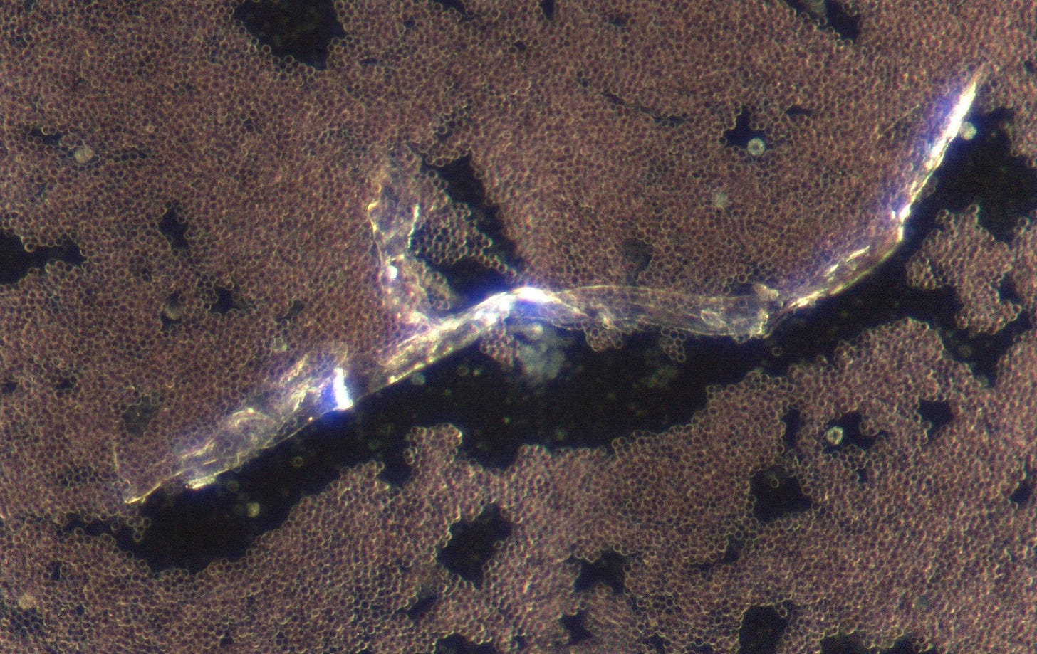
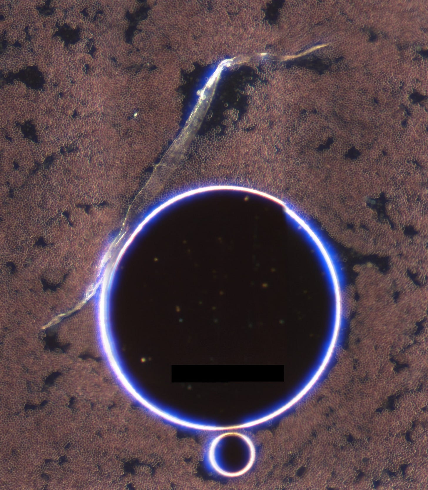
Here are two examples of what I found. Both are darkfield at approximately 40x magnification. Because the blood is mixed with EDTA and is 3 weeks old at this point this prevents comment with regard to red blood cells. Please disregard the bubble in the second picture. These are similar to structures commonly found in vaccinated and unvaccinated human blood (and the cows I have checked too).
With respect to the medications
First of all I looked at the ketamine and this seemed to be fine and it crystallised in a predictable fashion.
Here are a couple of videos that show this.
As soon as the medetomidine started drying I realised a familiar pattern. This looked similar to the dental anaesthetic except that it wasn’t preceded by the rainbow halo nor followed by the unusual cascade that I have documented in the last post.
The micelles that I had seen previously however were all to familiar as were the crystals that subsequently formed. The list of ingredients on the Zoetis supplied Safety Data Sheet mentioned only 4 ingredients 1 of which was water. Although medetomidine is a white crystalline structure I am confident that it does not look like the crystals I found.
Here is a video of the crystal formation.
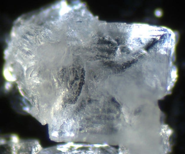
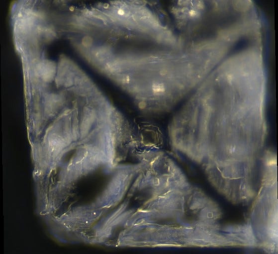
And here are some photos taken within the first 10 mins- Dark field x 40
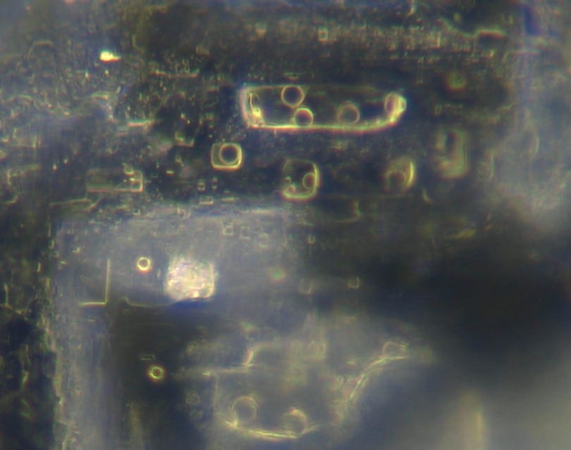
after 1-2 days I applied a fine oil to enhance the detail and these photos were taken subsequently of the same structures.
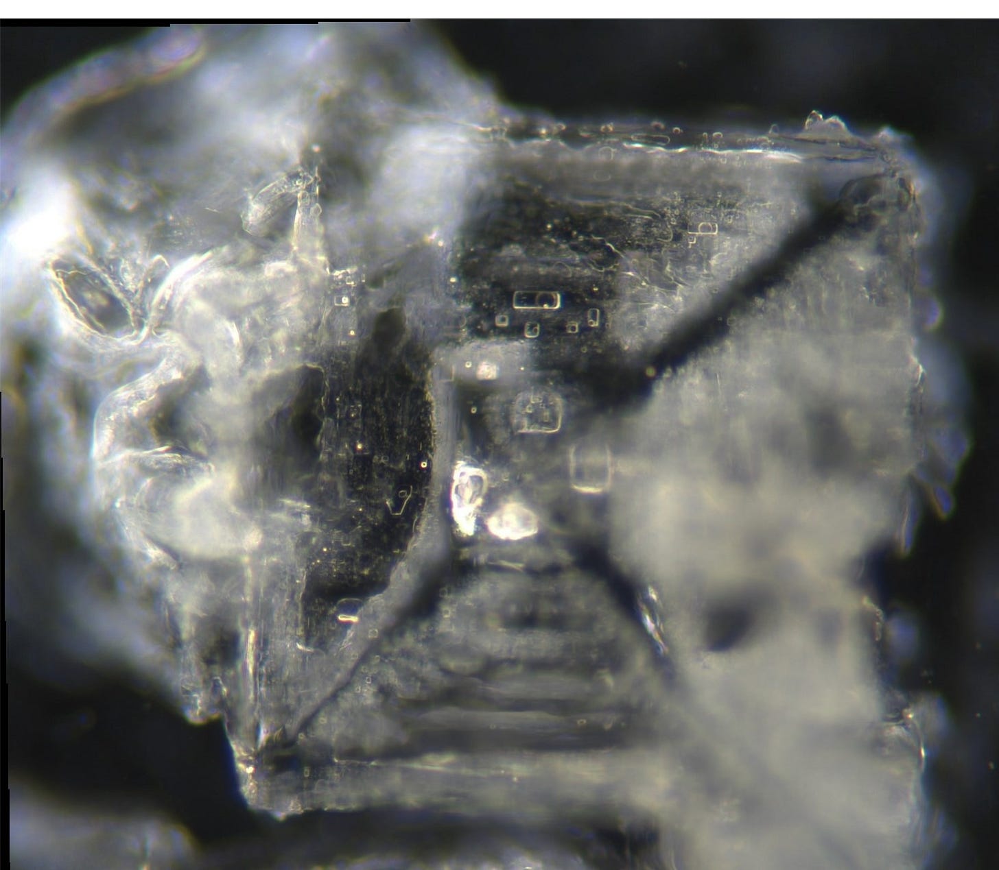
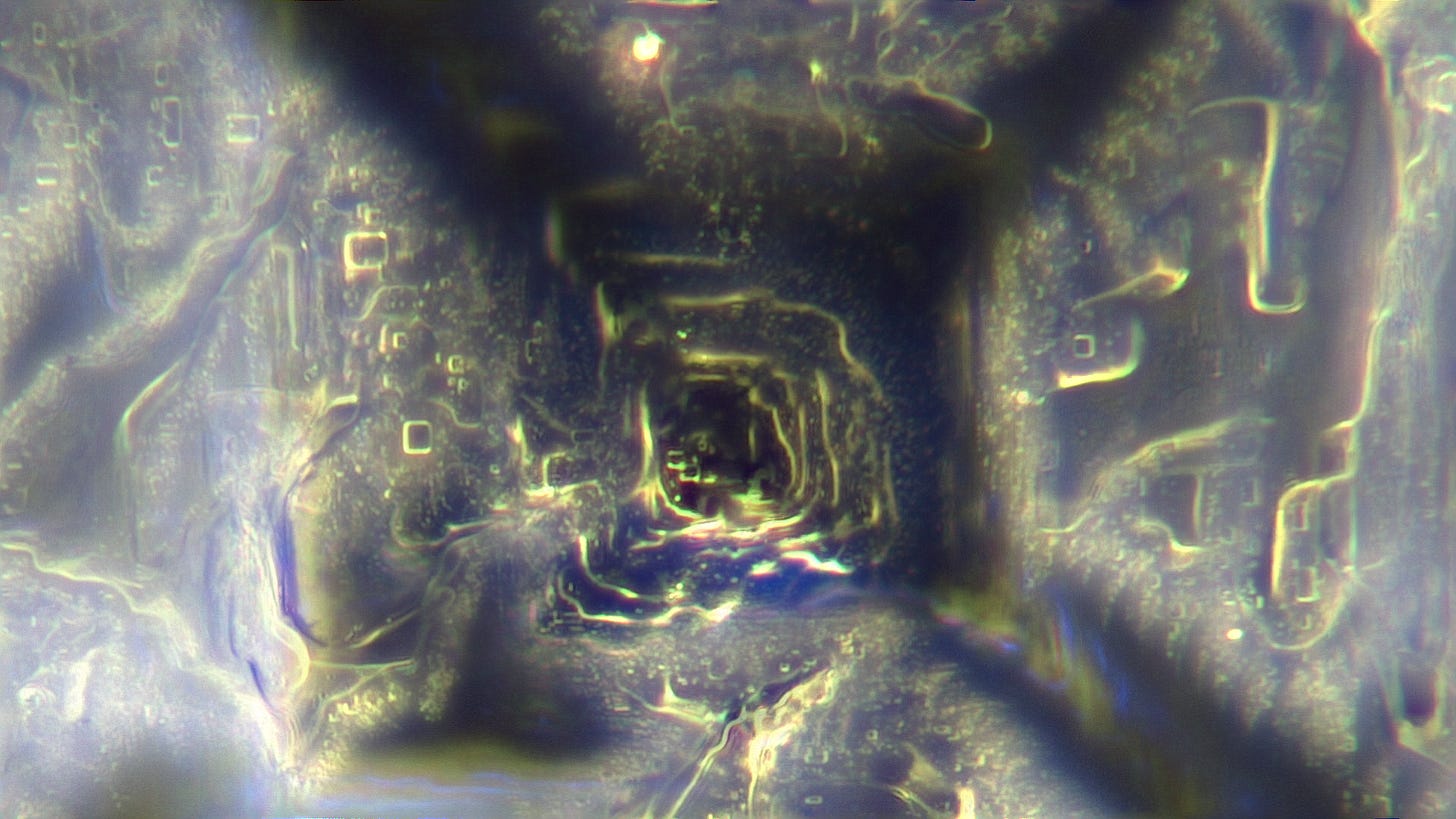
Detail of the above structures x 200 magnification
Another fresh chip x 40 and top left corner x 100
I have yet to see a ribbon attach to these latest crystals. I wonder whether the elaborate process observed in the anaesthetic makes the crystals electrically more attractive to the ribbons? The above structures seem similarly complex but who know whether they are functional in vitro or in vivo.
I offered recently to check all the injectables at a local medical clinic. An offer which was declined. “What if you are wrong?” Well I am 100% sure that I am not wrong but that in itself does not help the argument much. What if I am right? In my opinion electron microscopy will not reveal the detail under the top layer - I may be wrong about this. I am sure that eventual analysis will show materials that are not listed… I am not sure that this will help much either… We need to systematically look at everything we can, clearly…
These structures are technology.
Like the structures I have observed in the vaxxines and the local anaesthetic they become more organised with time and please compare the last image with the one above. I have videoed this area over 12 hours and captured this happening - I will put this on my website.
Please buy a t-shirt or two from the supporters store: https://nixonlab.myshopify.com/collections/all
and if you have a particular image you would like on a shirt please let me know and I will do my best.
Lastly, I will be offering twice weekly zoom meetings for my paying subscribers and I will vary the times. If you would like to join in you know what to do!
Have great weekend!
David







Great stuff David!
Here’s my thoughts:
The video of crystal formation (chip assembly) (12:36):
I’ll skip to the five minute mark:
@ 5:00 I believe we’re seeing a coiled up tubular construct (“Tube”) full of chip building material. A bit like a garden hose on the ground.
There appears to be a controller sphere floating inside the area bounded by the Tube. This controller sphere seems to be directing operations. There is also a smaller sphere.
@ 5:30 you can see both of these spheres appear to be photonically signalling to the Tube and probably each other.
@ 5:45 a third sphere comes zooming in from the right and attaches itself to the base of the Tube. I believe this sphere is the first of two that prepare the tube to begin chip construction. When it stops on the Tube it too becomes photonically active.
@ 6:15 you can see the larger controller sphere is flashing like a Christmas decoration.
@ 6.30 you see another small controller sphere come in from above the tube and also makes its way to the same place on the base of the Tube. When this one arrives and attaches the Tube instantly moves in a minor convulsion as it absorbs a smaller Tube of material that was in contact with its left side. Immediately after this the small controller sphere goes dark (mission accomplished?).
@ 6:45 the coiled Tube is slowly being moved into position for the chip building.
@ 7:15 there is increasing activity at the base of the Tube – things are about to get very busy.
@ 7:35 the large controller sphere has increased in size and is very photonically active.
@ 7:44 the large controller sphere goes dark (Mission accomplished?).
@ 8:00 there seems to be preparation (pale green and blue dots) taking place just to the right and base of the Tube.
To the left of the picture we see a totally separate chip suddenly spring into assembly from the edge of the sample – there appear to be two dark cables attached to the underside of it leading back down into the matrix. There’s a couple of bots tending to it whilst raw materials are fed into the chip.
@ 8:50 When chip construction commences under the Tube we see the Tube beginning to uncoil until there is an explosive uncoiling of the Tube. There is then rapid outflow of building material from the uncoiled Tube into the chip being assembled.
@ 9:00 There appear to be some structures with blue lights under the Matrix where the coiled Tube was located and further over to the right. If you fast forward and back from 7:20 to 9:03 you will see what appears to be a black cable from below (south of) the sample moving along the edge of the sample - it's movement seems to be coordinated with the rest of the activity occurring. You may have to switch off the light to see it clearly.
@ 9:05 you can watch construction of the chip with several bots in attendance – most inside the chip – but you can see them moving around inside. Incidentally the bots you can see are supervisor bots - the construction is being carried out by swarms of nanobots that are too small to individually resolve.
I believe the dark streaks we see appearing @ 9:20 under the chip and around the vicinity of the chip may be signs of these nanobot swarms. Similar black lines can also be seen leading into the other chips in this video.
@ 10.05 you can watch the appearance of a bot that I’ve called “Green Lantern” as it’s wearing a green light on its head presumably for photonic communication.
@ 10:45 the view switches to a chip that looks close to completion. There are some bots in attendance.
@ 10:50 we see a second chip making contact – a collision of chips! We can see several bots at the crash site exchanging particulars.
@ 11:30 the view moves to a close up of the crash site.
The Photos:
In the second photograph of 3 week old blood - that isn’t a bubble - in fact it’s a great example of a coiled Tube. If you go to the 10 o'clock position on its perimeter you will see that the outer coil of the Tube passes directly into the ribbon.
The rest of the photos appear to show shallow pyramid shaped chips with an open center that appears to show up to 10 levels within. They must assemble the four faces of the pyramids much like a babies skull is formed with gaps between the bones of the skull. These gaps in the chips appear to be able to be filled in if and when basic assembly is complete.