

The blood reaches new levels of tragic alteration.
The blood reaches new levels of tragic alteration.
More undeniably obvious nanotechnology processes obsessed in HD.


The recent findings in blood have dire implications for mankind as human forms. So much has changed in our blood that I wonder if it is actually fully reversible at this stage. We have learnt a lot about this horrifying technology, the people who claimed to actually be doing it to us, and the loss it causes.
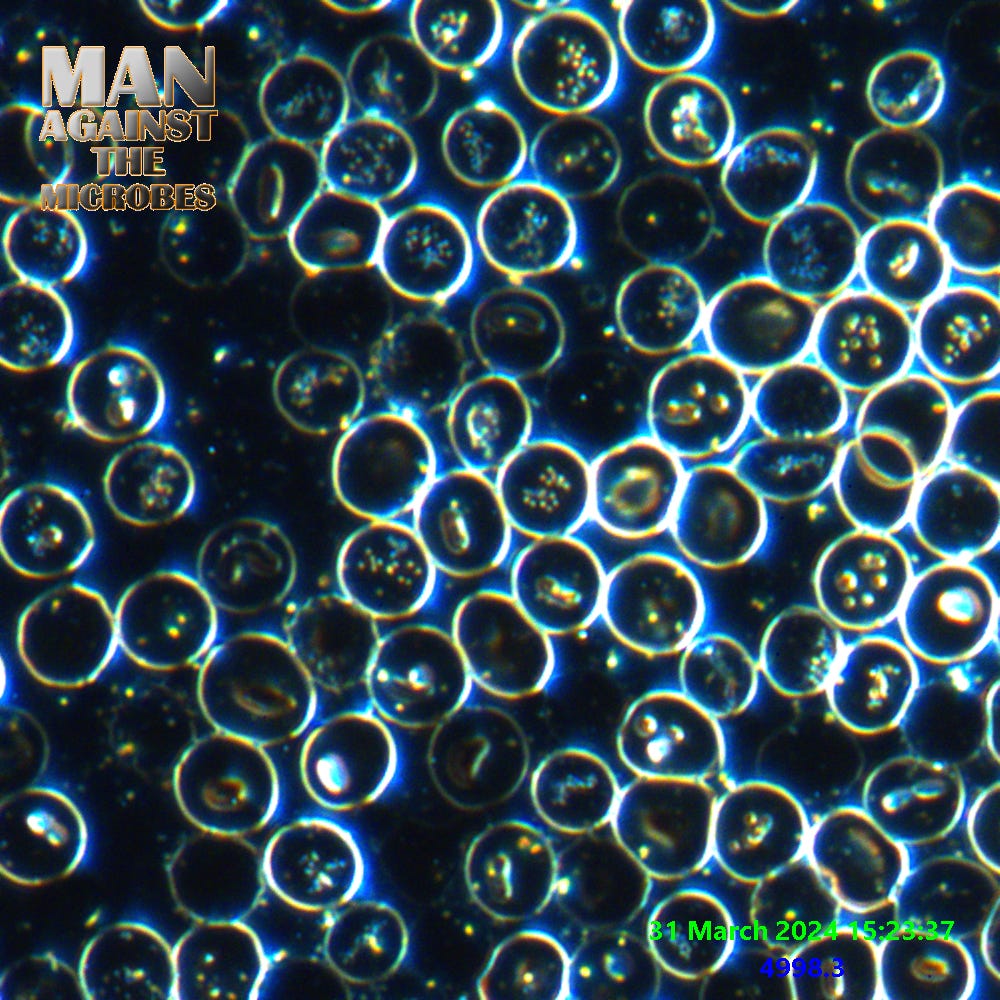
The above image is a collage of foreign cell inhabitants in our RBC’s. We have learnt during our endless research that these are just a few of the visual markers associated with colloid type nanotechnologies. These are larger forms that appear like clusters rather than colloidal molecules. There are many colloidal forms in the blood including expanded cell types. The whole package still seems to start with a hydrogel base and everything else is multiple pathways of hybridized nanotechnology all bolstered together.
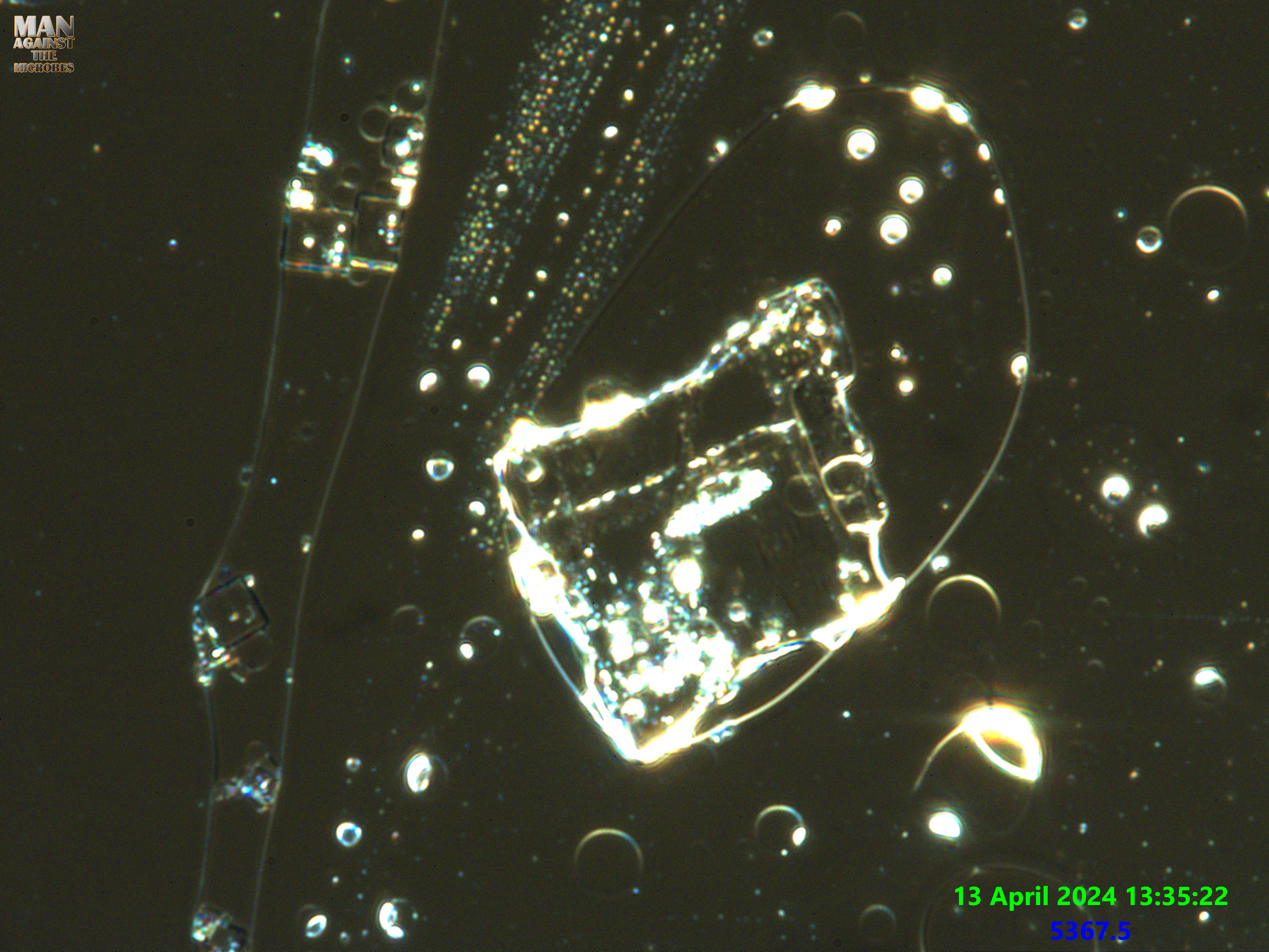
The image here shows a recent view of my own blood. The stages of development on this occasion are clearly more advanced. Due to the samples degrading it is unclear what these formations are purposed for at this stage. We recently noticed that the outer membrane of the RBC’s at this stage has become thick and polymer like.
The altered RBC membrane seems to have similarities to the constructor rings that many thought were bubbles in the blood. The membranes can be seen to consume material from the outer space and selectively draw in material to be processed within the cell. This is almost what all cells do in nature and this is why many stages of this technology use Protecell type forms to replicate and produce material at different ends. It works to form biological matter and can be manipulated to form other matter also.
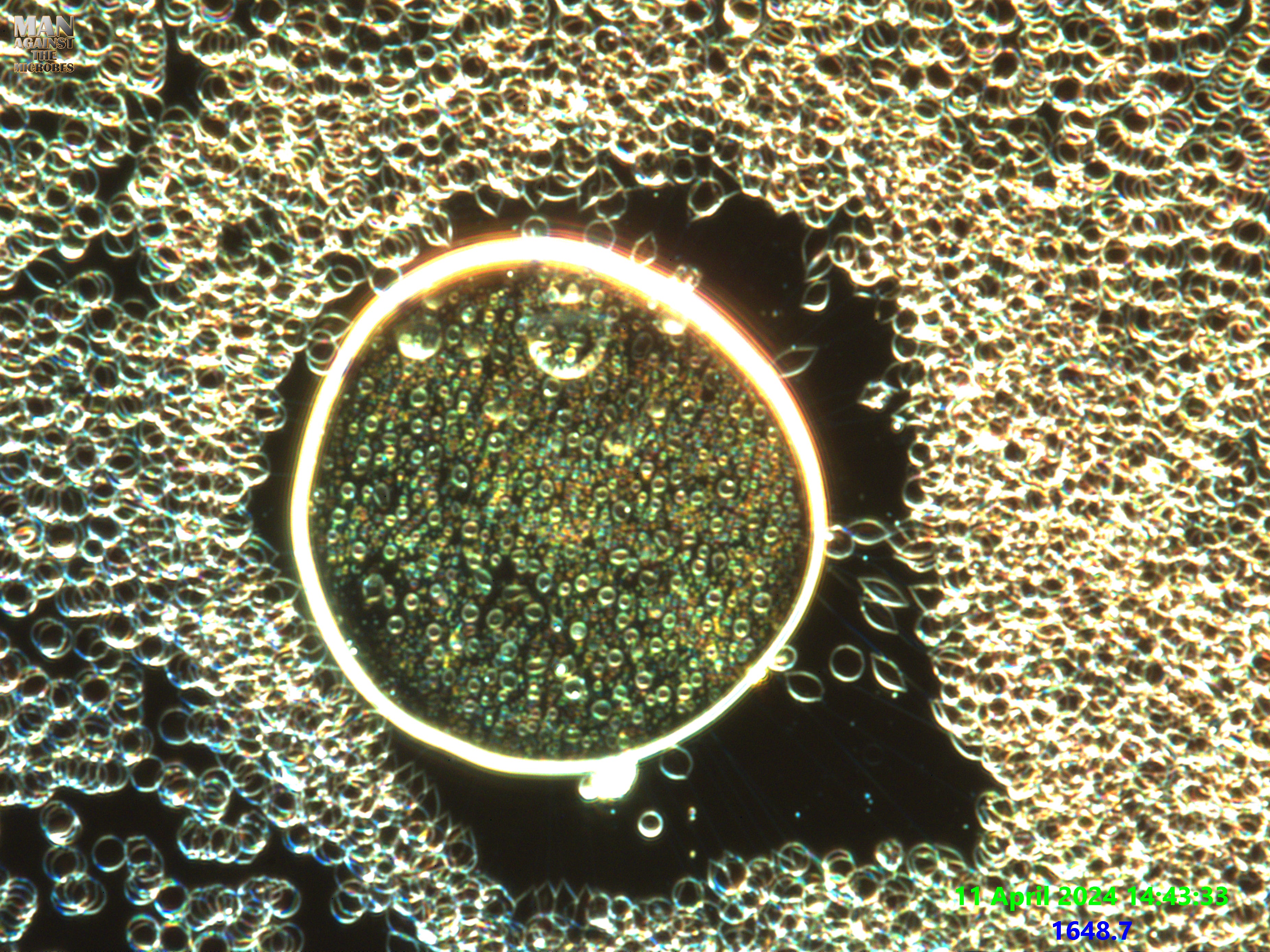
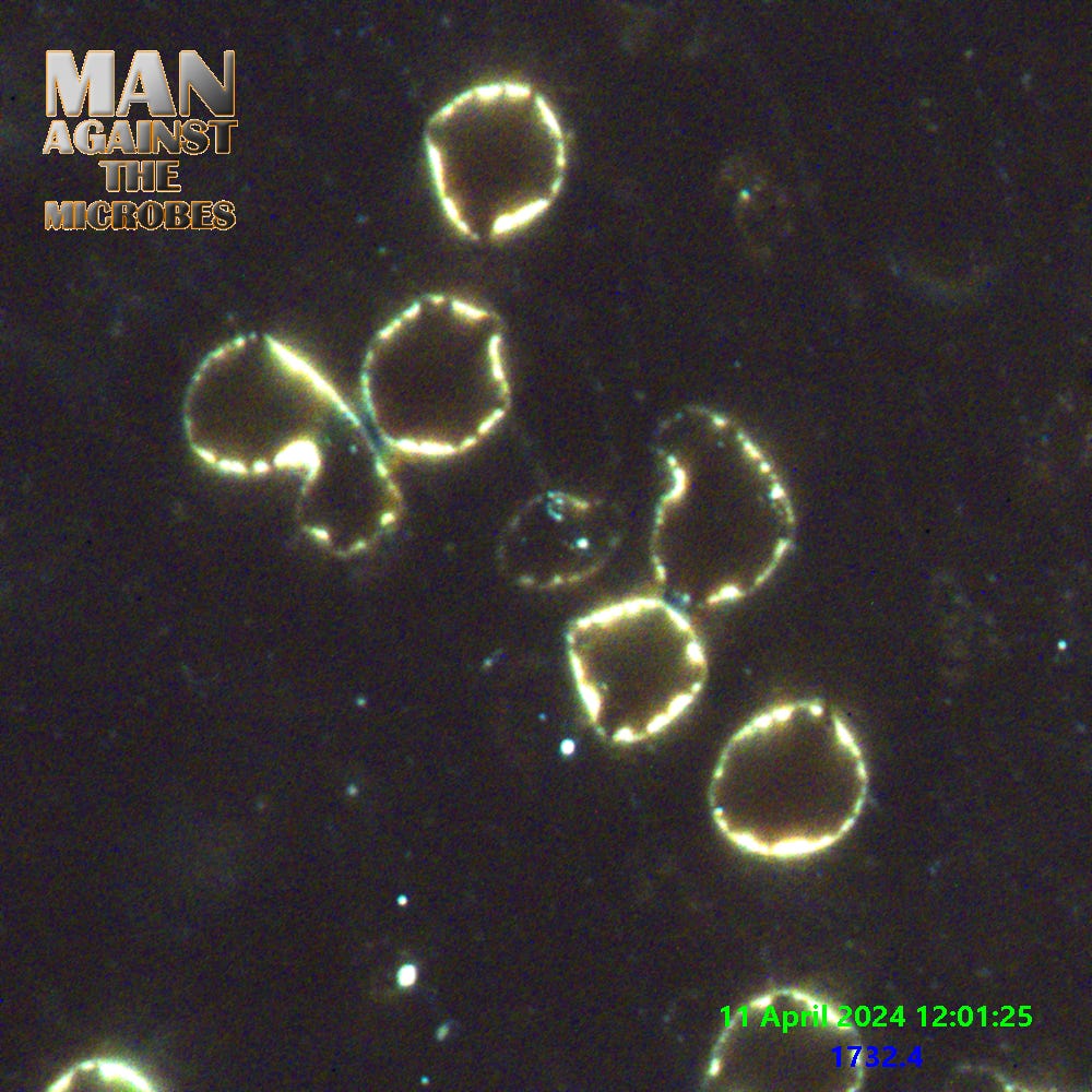
Above Is an example of lignocaine anesthetic, We use various injectable sources and Cov-19 swab products to examine this technology found in blood, water, and contaminated locations. They all exhibit almost the same complex material and morphological traits depending on the experiment conditions. In this image we see varying cell types which can be observed to differ more uniquely using dye’s. Colloidal molecules can clearly be seen with all their deceptive beauty as they trail up in a cluster of beads (top middle, rainbow colours).
The chip structure contains some types of these colloids and Nanoparticles which will undergo changes embedded in the salt like glass structure. Indeed in some images we can see trace like joins, moving structures, modular structures varying in colour, and then much more. Over all it looks very complex and unnaturally designed to assemble this way (not proven). Anyway the technical looking substrates are for another post since David and I have done a lot of work there too. David @
has spent a lot of time trying to observe these crystals and it is only just more recently we see these forms trying to express in some blood samples. The structures are not well understood but liquid and crystal embedded computing is obviously a topic we have been studying.
Our main concern here is that the blood of everyone I have seen on my high-end Leica microscope is looking like this or far worse as of a few weeks ago at least. It all seems rather consistent now. On a lower end scope like the average LBA would use these details are NOT CLEARLY VISABLE to this effect. The colour is lost and the resolution appears to show greenih lumps in RBC’s like nodules, and with faded dots in plasma instead of brightly fluorescing features. The image of a blood bubble cell is as I try describe them for now, a constructor cell with expanding colloids created inside which will produce various material such as colloidal cells, colloidal vesicles, and other cell structures. Some of these formed cells clearly have microfluidic systems inside as I have been seeing for nearly 7 or 8 months now at least. Below is an example of the microfluidics pumping away in a blood sample that had a COV-19 RAT test solution added and dyes. To someone who is not observant this is of course just evaporation :) Video is at 20x speed.
The work and images provided by
and are free to be used anywhere as long as reference is used and credit is given. Please also provide links to the source. This ensures we get help continuing our studies
The videos showing microfluidic systems (chemical reactors) are taken from blood, and other samples. We have seen these consistently. The tubes connect the vesicles and the colloidal formed sacks or cells are able it would seem to selectively allow material to pass the membranes so chemicals and particles can be mixed specifically in reactions and then transferred to the next stage. It is a modular factory that can be seen as a productive stage in all this horror. Jut one more piece in the complex puzzle. Slowly it all slots together. An in situ, self assembling microfluidic chemical reactor system.
There are endless mind blowing images and videos that we have not had time to share outside of closed research groups. In order to further our analysis we need help to afford the very expensive lab gear to provide us with the data.
The above structures sometimes will form from the colloidal structures in the constructor rings below. They also form on polymer substrates like the 2nd video up and this can be seen at the beginning of the clip when the whole hex shaped structure moves for a split second. The microfluidic structures will also form on the glass of the slide also. We have endless, unique cell structures observed and I personally feel mildly overwhelmed while getting all this info together in an orderly fashion with such little help and resources as we have so far. But it will get done !
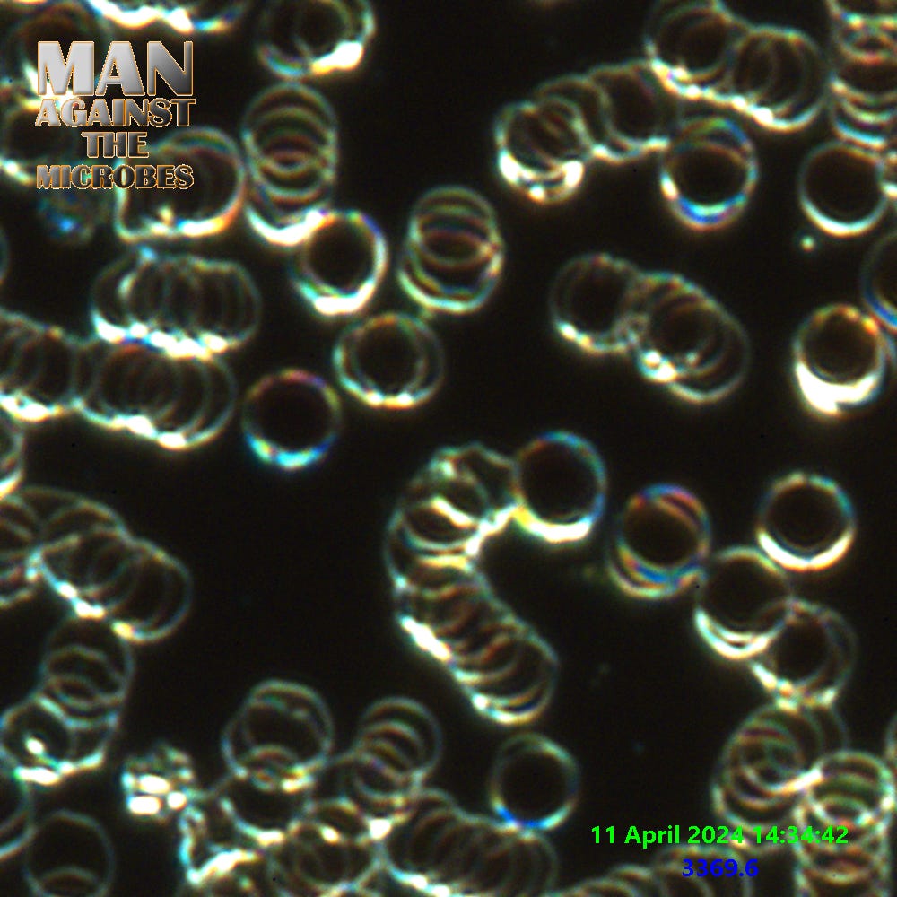
The ring structure does seem to harvest RBC’s. it even has strings pinned to each of these cells and the cells move in towards the structure before being converted into other products on the other side or the inside. Mind boggling to watch it over and over, but it happens and you can see it.

I can even see the material coming out of the membrane at very high magnification and with lots of optical tweaking on the scope to get everything just right. Below shows the colourful altered RBC membranes.
I hope that we have shown clearly enough over the last year that anyone touting this technology as being just graphene based without acknowledging that it is largely a hybrid scaffolding approach to nanotechnology roll out is really yanking your chain. I get tired of hearing people say “why don’t you mention the graphene?” and the reason I do not mention graphene all the time is for the same reason. I wonder why the Graphene crowd cannot see all the obvious stuff that the rest of us see, It clearly is not Graphene so much as polymers, hydrogel, coacervates, protecells, colloids, and NP’s we are seeing. Is that not bad enough, if not even worse than just Graphene alone? (There may well be some graphene involved!) but hey, lets talk about why the most obvious materials are being completely missed by the Graphene pushers.
We don’t fully have the order of everything in exact place yet and I wish not to misinform people by inferring more than i feel fairly certain about at this stage. We have a whole tonne of work to lace together and present carefully. But we really would feel much better getting that HPLC Liquid Chromatography going and a Raman microscope. Please help us reach our goals if you can. Those things cost about 100-150k or more used.
A huge THANK YOU to
(yet again) for being such a great contributor to our work with her kindness. So much love and hugs from us!Thank you to all who have helped or are considering helping. Thank you to the Micronaughts, The readers, supporters, other researchers, and my good friend
. Stay strong, stay human !