When I was at school I can remember handing in a sub-standard assignment and being chastised with “a bad worker blames his tools”. I am fairly sure this was due to some obviously poor excuse to explain my messy handwriting… (it’s got worse)
This is NOT that.
This is a big deal and I expect this is deliberately being done to distract from what is really going on. Let me explain.
Since the time of the vaccine roll out health practitioners that perform checks on live blood have noted abnormalities in the blood. This has included clumping of red blood cells so-called ‘rouleaux’, ribbon like structures and homogenous lumps of amorphous material which have been labelled as biofilm, protoplasts or crystals. I am still unsure what we are looking at. There has been a lot of attention to these abnormalities but not by mainstream media, obviously. Here is a short article I wrote last year: https://drdavidnixon.com/1/en/topic/what-s-in-the-blood. I wrote it as a short follow up to an article in the spectator:
Over the last 18 months the form and structure of the abnormalities has changed. At the end of March this year we held a small meeting of interested practitioners and over three days we took several hundred photos. A selection are below:
There are those that would dismiss these abnormalities as “fibre and dust”, which rolls of the tongue as easily as “salt and cholesterol” but is equally unhelpful. Dr Rima Laibow refers to them as Frequency Responsive Self Assembling Structures, (FRSAS), which is far more useful.
The pattern of abnormalities has changed over time. Initially these were only seen in the vaccinated but this quickly changed, within months, to include the unvaccinated. Clearly this implies either a shedding phenomenon or an alternative source other than the vaccine, or both. A taxonomy of what we are seeing is beyond the scope of this post however I will discuss further in the near future.
For some time a colleague has been convinced that there is significant contamination on the coverslips that we have been using. I thought he was mistaken but after spending a few hours dipping coverslips in and out of HCL acid, water and other solutions I am also convinced.
This is not just a matter of dirty coverslips such as can be bought cheaply online. A move that I came to regret after getting shards of glass in my fingers trying to clean them. Nor is this a result of dusty offices or poor technique.
The solution to having to spend time cleaning coverslips is to buy more expensive coverslips. The ones that I purchased recently are also made in China but they come in a sealed container and look perfect to the naked eye. We have also checked cover slips made in Germany which are even more pricy however they are also affected with what appears to be exactly the same contamination - go figure.
Just to clarify the cover slip (=cover glass) is the thin sheet of glass that is carefully (most times!) -placed on top of the sample which is sitting on the slide. This improves clarity and provides a uniform focal length. My colleague occasionally takes a small sample of blood which does not cover the whole face area of the cover slip. On occasion he noted the presence of structures either on the top of the coverslip or away from the blood sample. Simple washing with detergent of alcohol did not stop this problem. Hence the HCL acid. Washing with HCL acid did not sort the problem. Well not for the length of time that we used which was approximately 15-20 secs.
This is what a perfectly good-looking coverslip looked like after it has been in acid and cleaned in distilled water:
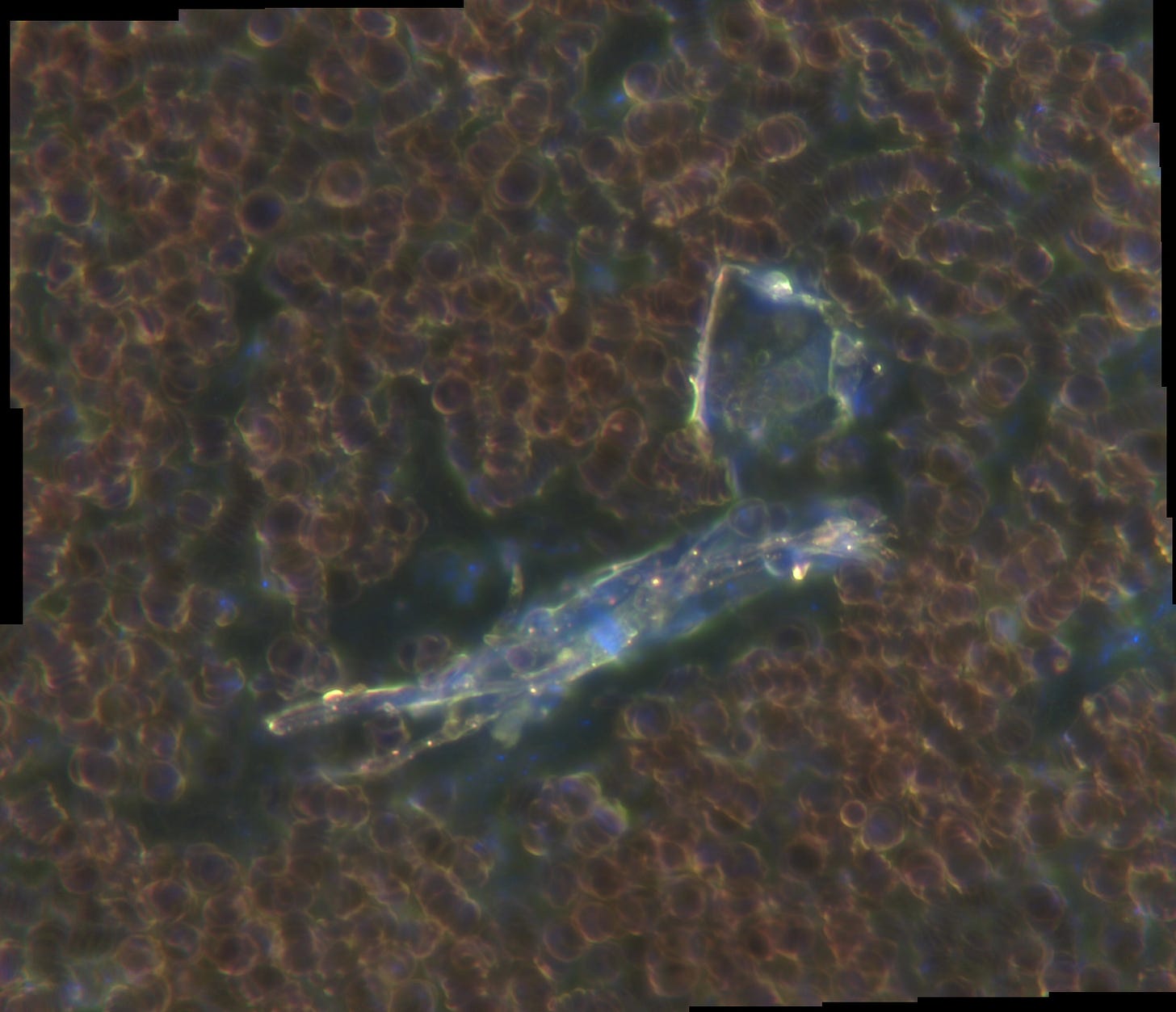
Here is an example of an unwashed coverslip carefully placed with suspected contamination face down. This one shows a typical structure and extensive rouleaux:
We found that when we used unwashed, 30 year old cover slips (my colleague has been doing this for a while) the blood looked like this:
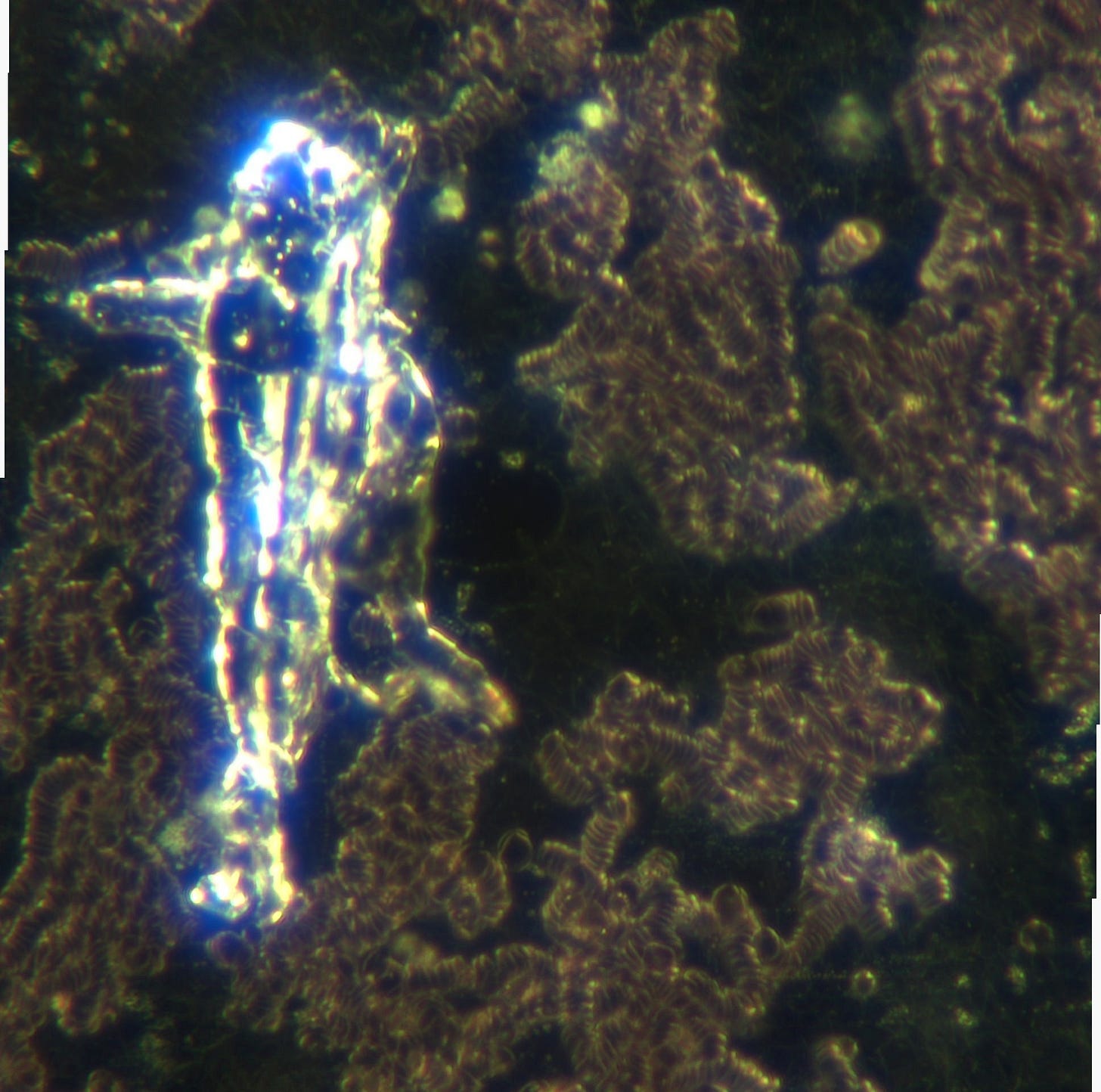
The remaining coverslips had various degrees of contamination despite been though an acid wash. This photo shows contamination remaining on top of the cover slip:
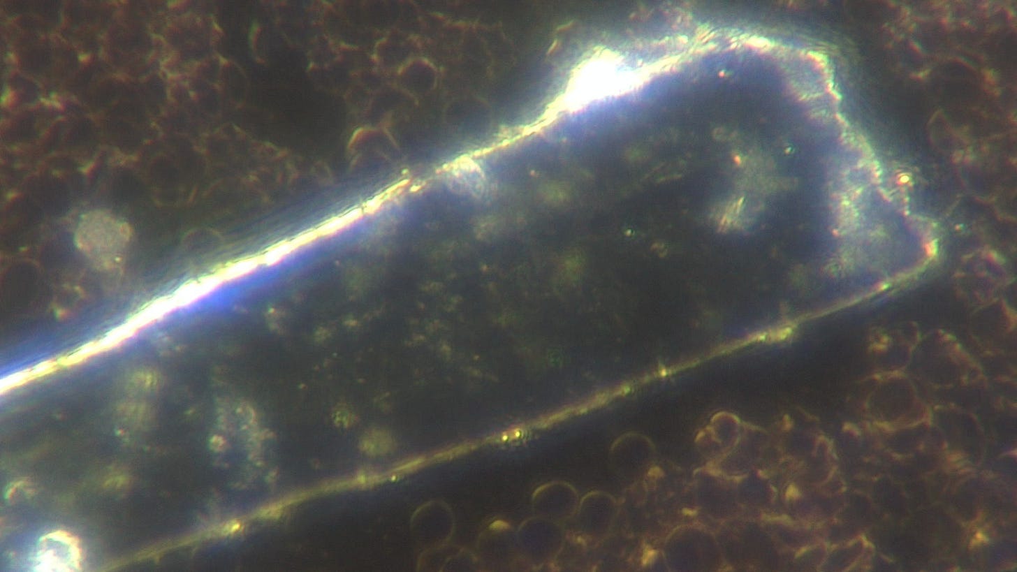
This one shows degraded contamination:
Over the last 3 and half years we have seen some unbelievable deception whether it be the “safe and effective” mantra, the mandates, the masks, the asymptomatic infection… the list goes on and on … and on.
I would like to add deliberate contamination of laboratory equipment.
This is a lot of trouble to go to to confuse and discredit - I think it is a deliberate distraction.
It reminds me of a TV documentary on WW2 I watched relatively recently (which means it feels like years ago but almost certainly wasn’t)… the comment was made that the “bigger the offensive: the greater the deception.”
https://nanocluster.mit.edu/publications
I am not for a moment implying that the Bawendi group are involved but this photo which depicts “Wizard Moungi” in Gollumesque pose does raise the question in my mind “FFS! What Could Possibly Go Wrong???”
It does make sense then to distract us with the “ribbons and the rouleaux” whilst the “dots” and the “lumps” are installed. This will take a bit of explaining so I will leave you with a bit of a teaser…
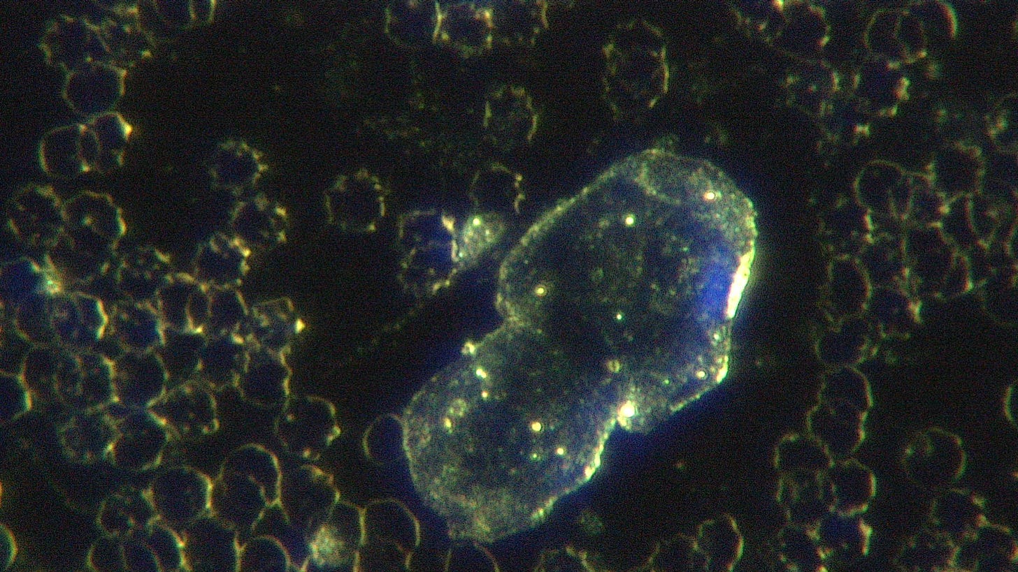
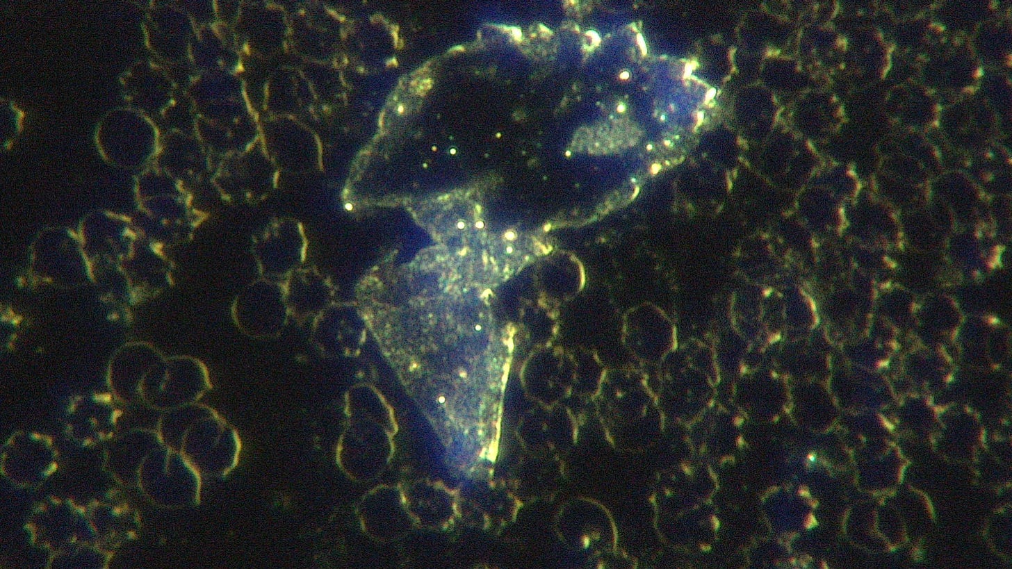
This is darkfield using 100x oil immersion objective producing 1000x magnification and shows a neutrophil which appears to be chasing a blue dancing dot. Blood was provided by a 28 year old male patient who had previously received 2 doses of Pfizer Covid-19 Vaccine. Significant adverse events included myocardial infarction, fatigue, asthma, blurred vision, rash, brain fog and EMR sensitivity. He had been fasting for 15 hours prior to the his sample been taken.
Back with more dots and lumps soon.
David
PS Elvis makes an appearance at 5:53








Thank you for this blog, Dr Nixon. As someone who hasn’t gotten to analyzing biological fluids yet until I exhaust analyzing injectable pharmaceuticals under a microscope (I am studying dental anesthetics at the moment), I understand the slip cover issue.
However, the fact that the structures (FRSAS) don’t appear immediately, but need at least 24 hours (maybe less) to assemble, deflect the dirty slipcover argument - to a point. I can envision the scenario when the coverslips are contaminated with smaller precursors of FRSAS that assemble once they have the opportunity to move toward each other using the liquid matrix of the sample studied.
I also want to support Dr Nixon’s sentiment “the greater the offense, the bigger the deception”. The rules of the game have changed, and people who are playing yesterday’s game, are not cognitively equipped to fighting today’s war.
And you can put away labeling something a conspiracy theory. This maneuver is no longer working, or valid, or carry any significant value. There is no denying that people worked on this technology together outside of the scrutiny of the public eye - that is your classic definition of conspiracy. So this labeling trick is moot. Might as well go put that back on the shelf, and engage in the conversation about what matters the most - what are we really facing here? And what is the ultimate purpose of this world-changing technology.
I'm going to call the blue dot Vlad The Blood Mozzie. 🧛
I reckon with my layman's degree in Street smartz cynicism, that "they" ( the Weasley Powers-That-Be's lackeys) have infected the supply chain of packaging of various kinds. I was buying Abbots ( a Vic brand) organic seeded sourdough bread but got worried about the plastic, which is thick and rubbery, so switched to Baker's Delight, non organic seeded sourdough because their plastic is the same as I remember bread plastic being. ( Considering getting a breadmaker to avoid this altogether. ) ( ..The appliance kind, not kidnapping a sleepy baker on his way to work)
I'm wondering if you shaved some plastic off the outside of that container the slides come in, what you'd find. Might be the container that contaminates them. Or if they come individually wrapped, the wrapper. ( No idea how to shave a plastic container, but putting it out there)
I even worry it's in small electric appliances so am in two minds about the ice cream maker for my brother's birthday ( for VEGAN ice cream! ...That's the point of buying it... Don't @ me 😂 ) 🍨
Also I found organic Camu Camu powder has 400 X the vitamin C of an orange. 🍊🍊🍊 For anyone that can't organise Vitamin C IV chelation, might help. ( Better than gmo corn ascorbic acid pills probably , which has the gene that allows them to pour infinite amounts of glyphosate on the corn, without killing the corn according to RFK Jnr recently.) .
Night from Australia 😴 🐈 🐾
I write a lot. I go now. 😬