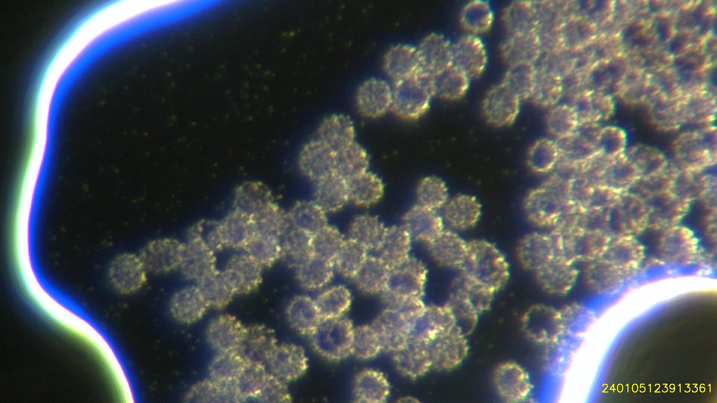
During a live blood check one other area I focus on is the edges of the sample and the interaction between the particles and the red blood cells under higher magnification.
I subsequently move into the more central area to observe how the neutrophils are coping with the particles.
Some peoples red blood cells appear to cope well with the hydrogel and the particles
whilst others don’t:
There is evidence from Karl C. that these represent particles on or in the red blood cells and he has some stunning images that show this.
Without doubt the fitter the person the least processed food they eat the better the cells cope.
This blood belongs to an ex-Olympic swimmer who remains in excellent health:
As you can see from this video using a 4x objective (40x magnification). As the red blood cells move towards the hydrogel margin the margin moves further away.
Here another couple of examples of this:
10x objective: (100x magnification)
20x objective (200x magnification)
40x objective (400x magnification)
Here are some particles and grossly affected red blood cells a little further from the edge of the sample:
100x oil objective (1000x magnification) showing deteriorating red blood cells and perfect synthetic cells of different sizes:
Three neutrophils seen using the 40x objective (400x magnification) showing large numbers of particles within the cytoplasm of the cell:
Neutrophil at 1000x magnification showing particles inside the cytoplasm:
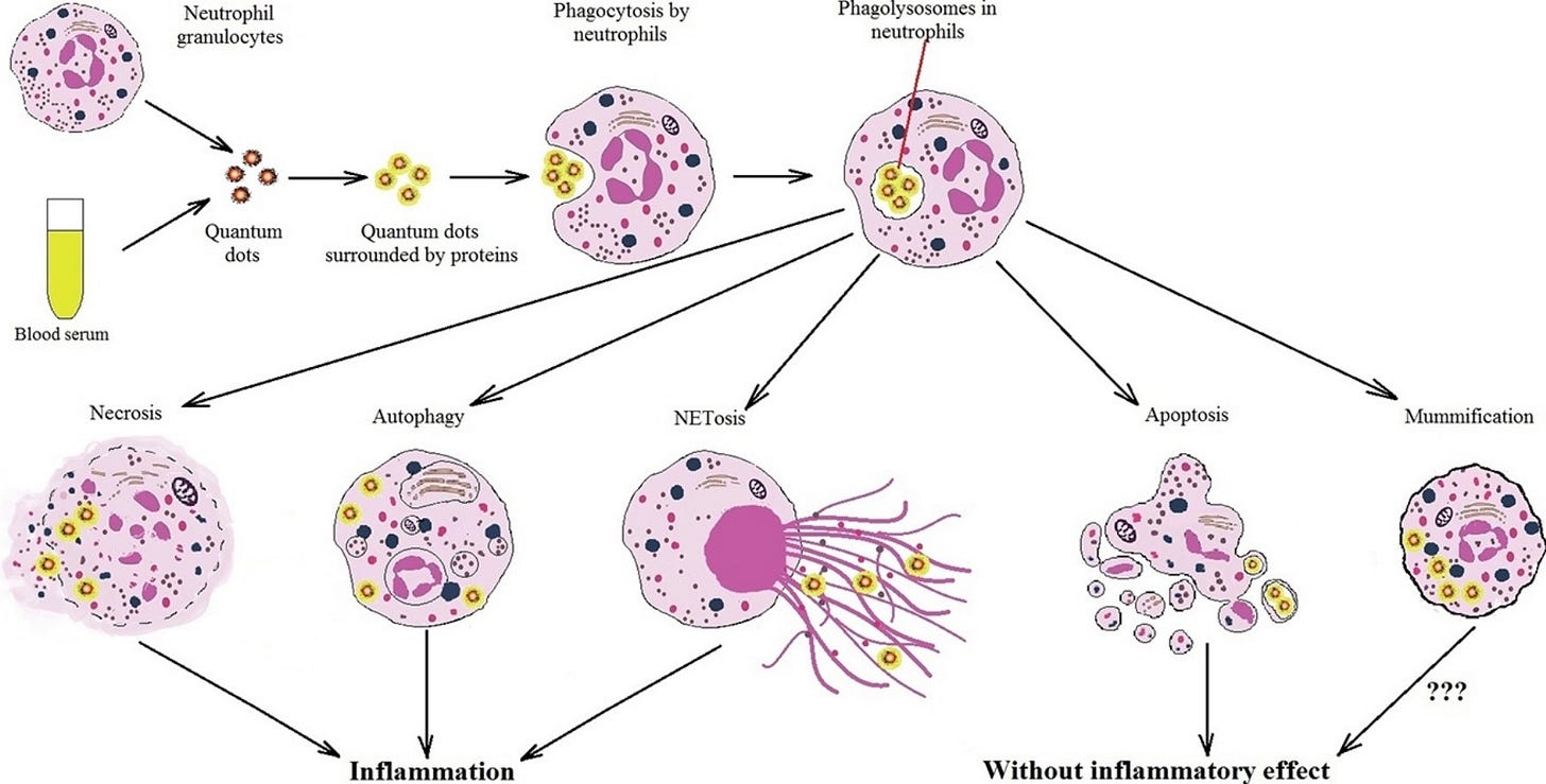
Here is a cartoon from a recent paper illustrating quantum dots been taken up by a neutrophil and thank you to a colleague for finding this:
I think in ‘real life’ it probably looks a bit like the videos above… ;-)
And this is the reference: https://www.sciencedirect.com/science/article/abs/pii/S096843281730450X
I will write more about the particles in the next instalment.
Ciao
David
And thank you for the support!


God Bless you David and crew for your amazing research into this topic! You are a blessing to Humanity for exploring truth and sharing it with us all.
Dr Nixon your last live video looks like a neutrophil engaged in phagocytosis.