Nano makes micro in an influenza vaccine made in Australia
Crystal-Fibre Assemblies and Circle-Rectangle Motifs amongst other interesting findings
The next time an expert proclaims that there is no nanotechnology in our medications ask them if they have looked at sessile droplet evaporation under dark field. If they haven’t (all of them so far) then please inform them that you won’t accept their opinion until they do! Politely.
I believe we are seeing highly sophisticated nanotechnology that builds microtechnology.
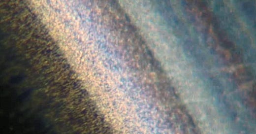
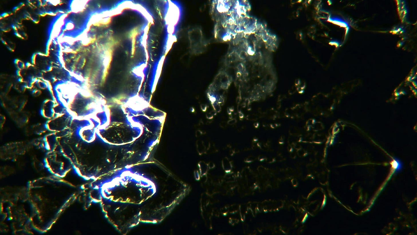
This photo above is of the sessile droplet evaporation process in Lignospan Special by Septodont. Septodont also state on their website that their products contain special molecules and they are safe and effective so I am sure there is nothing to be concerned about. Phew. My understanding is that the colours are due to the interference pattern from the nanoparticles and the light. We can’t resolve the individual nanoparticles. However they are present as colloidal particles in suspension and these self-assemble into larger colloidal particles which we can see. And these particles self-assembly into larger complex crystals which we can study in detail. It is not hard to have a look yourself or find somebody else that is looking.
The process of sessile droplet evaporation (SDE) is well studied. Please see Zang et al (2019) for more details. The physics and maths behind the drying of a drop on a hard surface is well understood and is the basis of ink jet printers and touch screens. The evaporate process causes all sorts of interesting stuff to happen. Capillary flows, mixing, concentration of particles...
One of the most interesting phenomenon is the Crystal-Fiber Assembly. Here is one in Pfizer Comirnaty:
Here is a Septodont special at 5 months old showing stability of the structure:
and here is one in an influenza vaccine - Afluria Quad:
and here is a hypothetical mechanism:
Nanoscale particles aggregate along capillary flows and drying fronts.
Electromagnetic or electrical fields guide and stabilize the formation into a fibre structure.
Self-assembling frameworks (potentially involving DNA origami or peptide scaffolds) facilitate the fibre's growth and attachment to the crystal.
Metals and other elements enhance structural integrity, leading to persistent and complex morphologies.
The fibre's formation likely results from a convergence of physical evaporation dynamics and self assembly principles. Apparently.
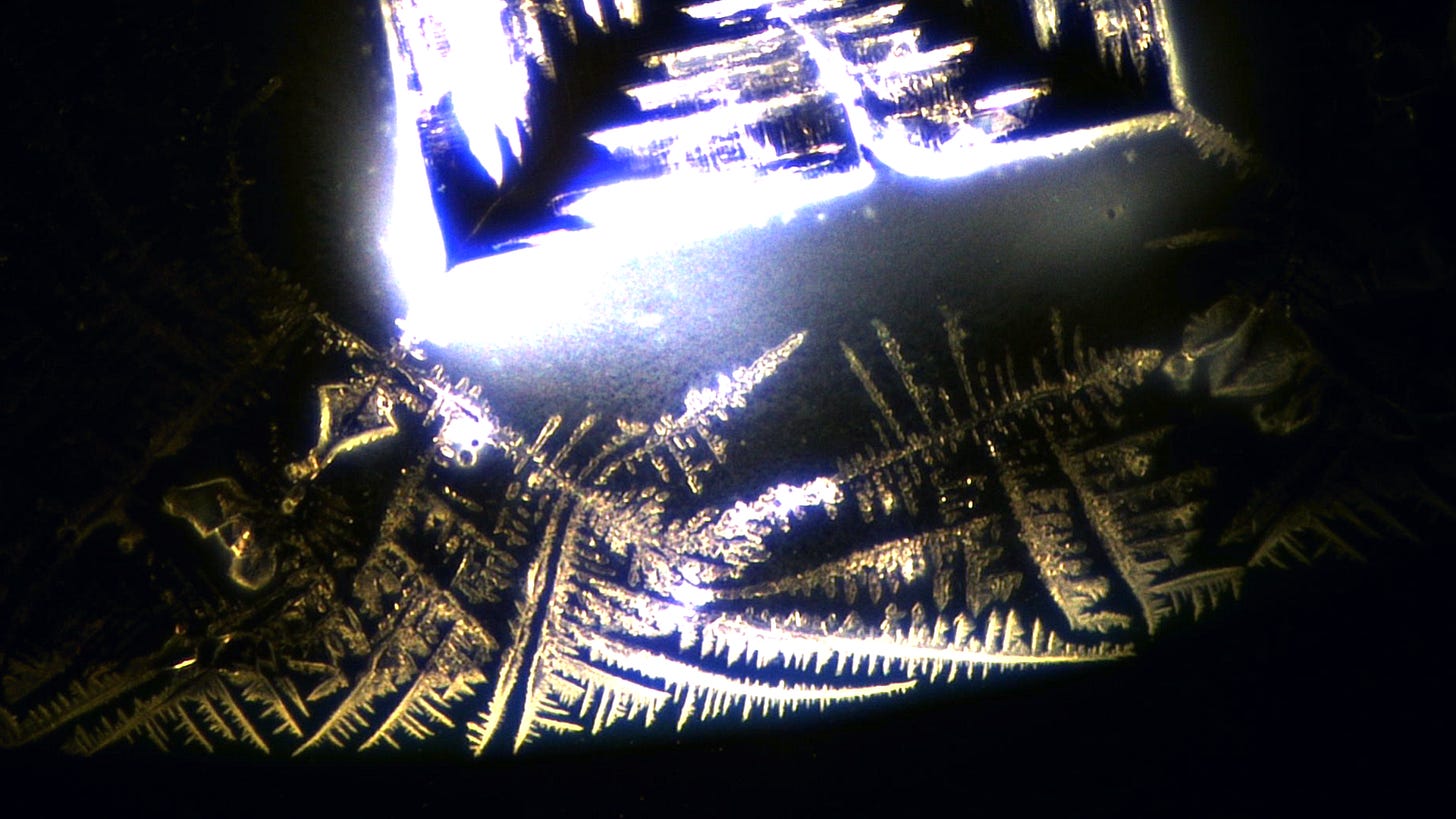
Another indication of synthetic crystallisation is the Circle-Rectangle Motif (CRM), which often appears in a fractal-like manner across different scales. When Mat Taylor and I examined Pfizer samples two years ago, we referred to the circular features as "donuts" due to their appearance as black rings under bright field microscopy and their high reflectivity under dark field. Over time, the rectangular elements of this motif also exhibit increasing geometric precision. This crystal is from a Pfizer sample at 2 months old:
This week I spent some time looking at a quadrivalent influenza vaccine.
It was in fact this one, recently provided by Rob - cheers mate. Afluria Quad, brought to you by Seqirus and made in Melbourne.
This image shows the initial drop drying on the slide at 100x magnification, using dark field microscopy with a polarising filter and likely a slight green digital filter. Dense colloidal material is visible within the drop, accumulating at the edge. The presence of such dense colloids is puzzling, especially given that this is the third flu vaccine I have examined this year, and these colloids are by far the most prolific
This was after the first evaporative process. One crystal formed with some interesting crystallisation patterns in the background:
What we see when the crystals dissolve is often very revealing and this time there were a number of surprises…
The image below displays a network of cells and fibres reminiscent of structures previously observed in blood samples. Such formations are neither normal nor expected. They suggest a remarkable degree of self-assembly and features indicative of synthetic design within the vaccine components — in my opinion, of course.
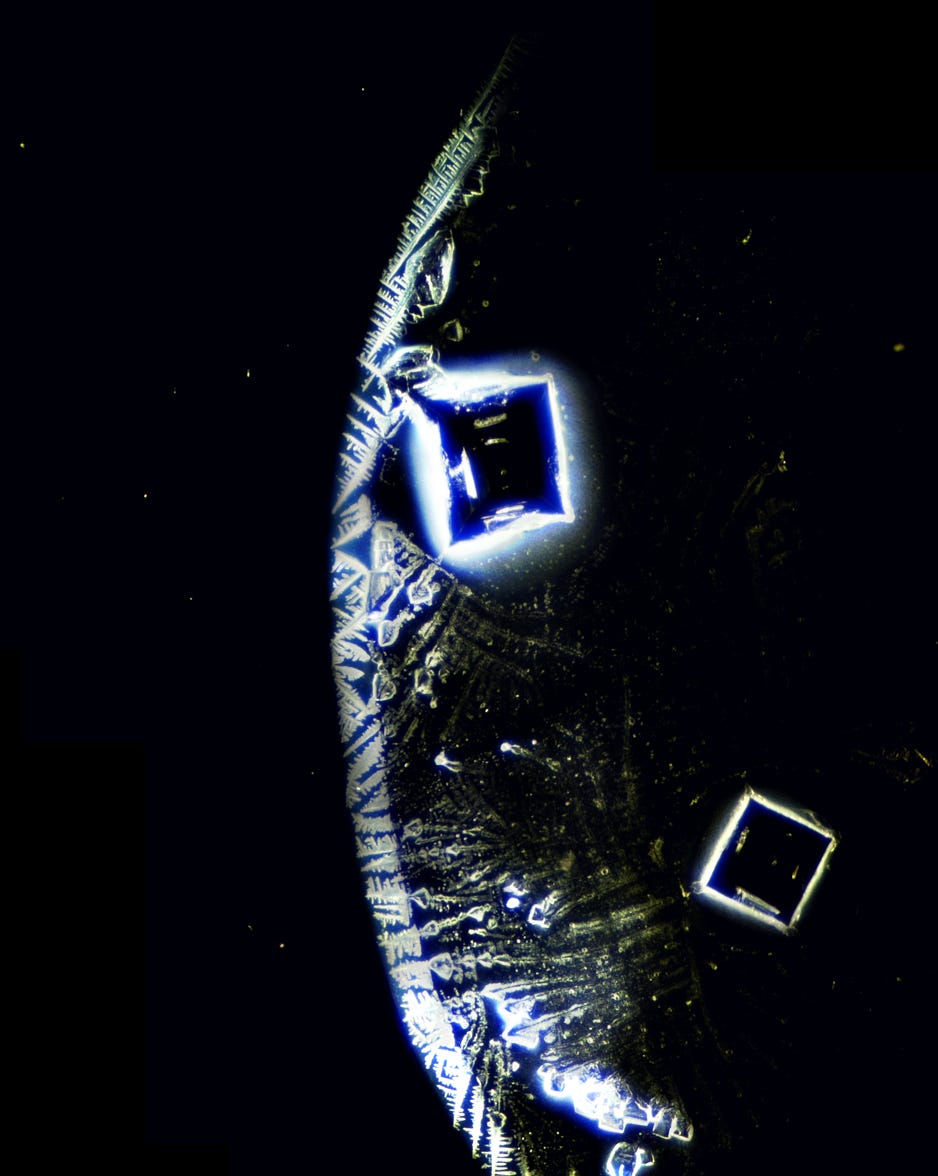
There was also this unusual looking membrane which I have not seen previously. Here is a close up of the edge at 200x magnification .
Here is a structure that looks similar and is the same size as structures that we see in This structure resembles those seen in blood samples and is of a comparable size. Notably, I have not encountered such formations in a flu shot before.
Here is a short one minute real-time video of a crystal forming from the second SDE process:
Thanks to Freedom Warrior Woman who coined the term pizza to describe the appearance of the dried drop. The large crystal is the one in the video above and the membrane is shown with the red arrow.
Here is a close up of the crystal:
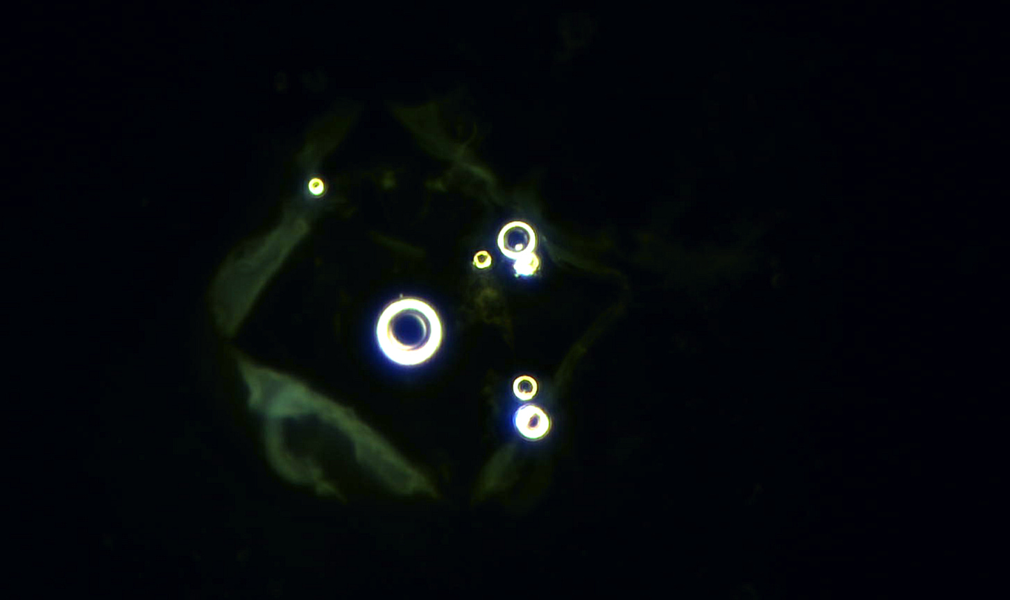
The central bubble shown here is not an air bubble but likely an accumulation of nanoparticles concentrated during the evaporation process. I first observed this phenomenon in dental anesthetic, but it has since appeared in multiple medications and even heavily contaminated rainwater. When the crystal dissolves, the bubble forms a ring structure, with the largest ring typically surrounding the central bubble. This image is a close-up of these ring formations captured during the third sessile droplet evaporation (SDE) process.:
And here is a close up a few minutes later when the small rings had disappeared.
Note the membrane around the crystal - is this a protein scaffold to facilitate self-assembly?
Here is a 4min real-time video of the third crystallisation:
Here is the third pizza. As you can clearly see the crystal at 9 o’clock is very similar to the crystal in the second pizza.. I would say the chances of this happening randomly are virtually zero. Interesting.
You can also see that a number of the other crystals have a bubble on top.
Here is a photo-stitch at 200x of the uppermost crystal.
Here is a close up of the triangular crystal at the bottom which might be just salt but it will contain extra material too I am sure.
Here is a video of that crystal dissolving:
It is a shorter 1min video “It’s always faster to blow things up” - Karl C.
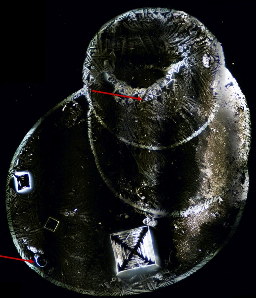
The image below is a photo-stitch using a 25x objective and a polarising filter above the light source and a modest digital filter. The large crystal at the bottom is visible to the naked eye (just), I think in the order of 0.5mm x 0.5mm. I added a small drop of C60 oil to the slide as I have found this improves clarity. You can see the fibre bottom arrow and the smaller crystal top arrow has a central crystal-rectangle motif which you can see in this image. It is the final super supreme pizza:
Here is a close up of the upper crystal:
and this is how it looked 12 hours before:
So you can see that it has changed and become more geometric - which they always do, in my experience. Here is a photo-stitch of the large crystal which is quite spectacular, followed by a few close up shots.
And here are a few other images from the slide:
Is this a protein scaffold around this crystal?
Here is a close up at 200x magnification of the fibre:
-I am sure that there are a number of live blood analysts who have seen these in blood samples. I think they are forming on the slide and Karl C has video of one rapidly forming so I don’t think is an unreasonable suggestion. This one is from a post I did on fibres and gel structures in the blood earlier in the year.
So the technology is in our medications and it is appearing in our blood…hmmm
Here are a few more close ups of the slide. I don’t think that the bubbles that regularly appear on top of the crystals are ‘normal’. I understand that there is often surfactants and other things in these medications but this happens far too often to not have a bioengineered aspect, in my opinion.
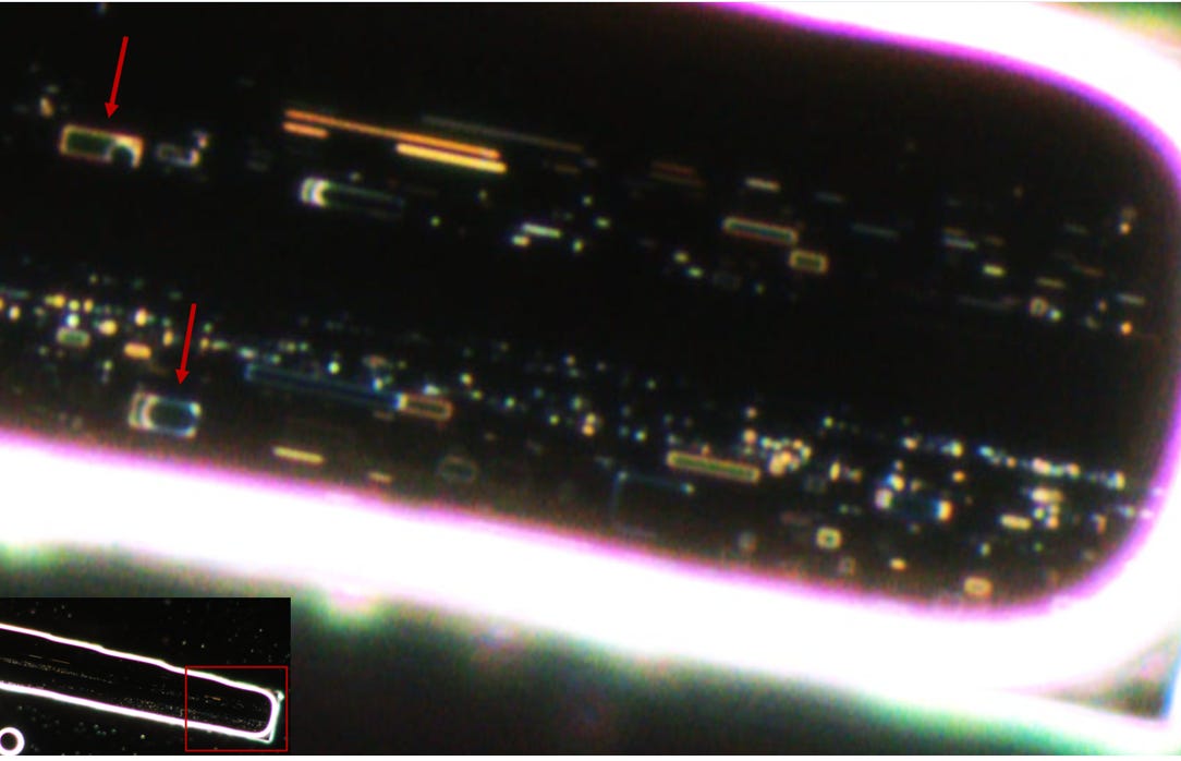
Lastly, the sessile droplet evaporation process happens under a coverslip too although it is a slower process. Thank you to Louise C. for providing this image. This is Citanest dental anaesthetic under a coverslip at 400x magnification. Arrows indicate circle-rectangle motif’s. I have included it here to illustrate how spectacular the technology can be… for more images please click on the image to go to my previous post.
Just waiting on a bit of feedback on my paper on dental anaesthetics but it is not far away.
Have a great weekend!
David
All assistance much appreciated! (Coffees are now reduced in price!)












I love this whole style of 'laid back' approach to telling this story here David, it flowed perfectly and you staged it so well with animations of perfectly crystal clear stunning captured.
Love this style of reporting, well done. More please✌️
Thank you David. These findings need to be exposed